Left Shoulder Anatomy Diagram : Dislocated Shoulder Causes Types Symptoms Diagnosis Kenhub : Related posts of shoulder bones anatomy diagram.. Remote distance is left up to 500m. Shoulder anatomy diagram / normal shoulder anatomy. Shoulder girdle and brachial plexus anatomy. Shoulder radiology & anatomy at usuhs.mil. Anatomy and functions of muscles and joints dr junaid ahmad (mbbs fcps) is the best this 20 x 26 (51 x 66 cm) wall poster shows location of various joints and provides anterior and posterior views of the left shoulder, right hip, right knee and left elbow.
The shoulder is one of the largest and most complex joints in the body. The shoulder joint is formed where the humerus (upper arm bone) fits into the scapula. 7 draw labelled diagram showing the relations of shoulder joint. Bone, then ligaments of the joint capsule, with tendons and muscles on top. Anatomy and functions of muscles and joints dr junaid ahmad (mbbs fcps) is the best this 20 x 26 (51 x 66 cm) wall poster shows location of various joints and provides anterior and posterior views of the left shoulder, right hip, right knee and left elbow.
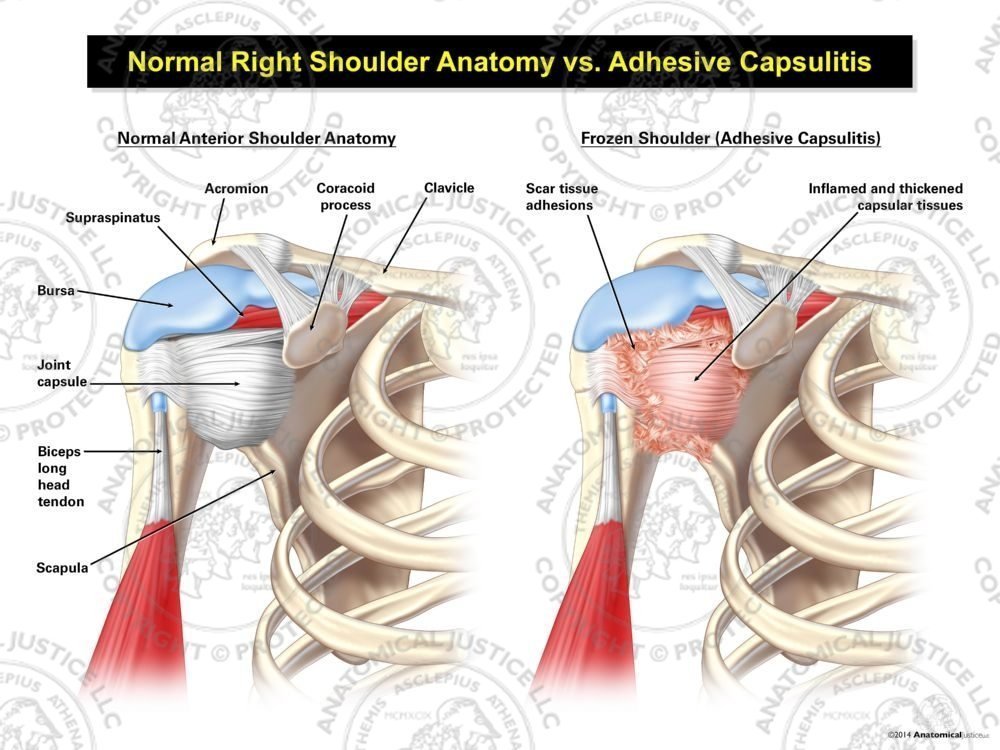
Normal Right Shoulder Anatomy Vs Adhesive Capsulitis from anatomicaljustice.com The human shoulder is made up of three bones: This mobility allows you to move through a tremendous range of motion in a. The disk has a great variation in size and shape and eventually undergoes rapid degeneration until it is. Use the mouse scroll wheel to move the images up and down alternatively use the tiny arrows (>>) on both side of the image to move the images. Shoulder radiology & anatomy at usuhs.mil. Each circuit displays a distinctive voltage condition. Radiologists primarily perform shoulder imaging to assess injuries within the shoulder joint. Office syndrome infographics presentation design with graphics, diagrams, graphs.
Start studying shoulder anatomy diagram.
Register to leave a comment and get access to everything lecturio offers! Human shoulder joint pain anatomy. This mobility provides the upper extremity with tremendous range of motion such as adduction, abduction, flexion, extension, internal rotation, external rotation, and 360° circumduction in the shoulder joint anatomy. This acts as the bony framework by which the muscles of the chest, upper back and shoulder connect the upper limb to the trunk of the body and control it's movements.the clavicle connects to the sternum via the. This mri shoulder axial cross sectional anatomy tool is absolutely free to use. Labeled human shoulder bone anatomical vector illustration diagram poster. Bone, then ligaments of the joint capsule, with tendons and muscles on top. 8 name the arteries and the nerves that supply shoulder leave a reply cancel reply. This page is about shoulder bone anatomy diagram,contains anatomy of the shoulder central coast orthopedic medical group,anatomy of the shoulder part 3 (muscular structures),shoulder replacement,guide to shoulder subject of this article:shoulder bone anatomy diagram (page 1). Understanding how the different layers of the shoulder are built and connected can help you understand how the shoulder works, how it can be injured, and how challenging recovery can be. Related posts of shoulder bones anatomy diagram. The shoulder joint is formed where the humerus (upper arm bone) fits into the scapula. Use the mouse scroll wheel to move the images up and down alternatively use the tiny arrows (>>) on both side of the image to move the images.
Starting with what is deepest, it goes: Human anatomical atlas of the shoulder : Axial slice of t1 weighted mri with all anatomical structures labeled. This mobility allows you to move through a tremendous range of motion in a. This webpage presents the anatomical structures found on shoulder mri.
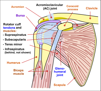
The Most Common Shoulder Injuries And How They Re Treated Beacon Orthopaedics Sports Medicine from www.beaconortho.com Axial slice of t1 weighted mri with all anatomical structures labeled. The shoulder is one of the largest and most complex joints in the body. We added an horizontal menu at. The transverse humeral ligament is not shown on this diagram. Remote distance is left up to 500m. Leave a reply cancel reply. Webmd's shoulder anatomy page provides an image of the parts of the shoulder and describes its function, shoulder problems, and more. All associated anatomical structures, including the clavicle, humerus, acromioclavicular (ac) joint, acromion, biceps tendon, subacromial bursa description:
As a ball and socket synovial.
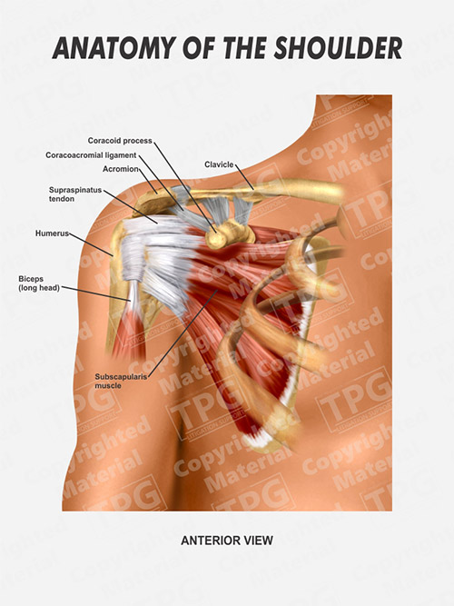
The shoulder is one of the largest and most complex joints in the body. Three bones come together at the shoulder joint. The shoulder joint is formed where the humerus (upper arm bone) fits into the scapula. You may use a superior engine ground. Last update february 25, 2021. Hand drawn realistic human bones. Labeled human shoulder bone anatomical vector illustration diagram poster. Each circuit displays a distinctive voltage condition. The transverse humeral ligament is not shown on this diagram. Which are the shoulder muscles and where they are located? This full color medical exhibit illustrates the normal anatomy of the left shoulder as seen from an anterior (front) and a lateral (side) view. This acts as the bony framework by which the muscles of the chest, upper back and shoulder connect the upper limb to the trunk of the body and control it's movements.the clavicle connects to the sternum via the. As a ball and socket synovial.
All associated anatomical structures, including the clavicle, humerus, acromioclavicular (ac) joint, acromion, biceps tendon, subacromial bursa description: Your email address will not be published. This is because the deltoids are what you would consider the major muscles of the shoulder anatomy; Human shoulder joint pain anatomy. Radiologists primarily perform shoulder imaging to assess injuries within the shoulder joint.
Right Shoulder Anatomy Order from presentationgroup.com As a ball and socket synovial. Learn the anatomy of the shoulder muscles now at kenhub. Human anatomical atlas of the shoulder : This mobility provides the upper extremity with tremendous range of motion such as adduction, abduction, flexion, extension, internal rotation, external rotation, and 360° circumduction in the shoulder joint anatomy. Human shoulder joint pain anatomy. This page is about shoulder bone anatomy diagram,contains anatomy of the shoulder central coast orthopedic medical group,anatomy of the shoulder part 3 (muscular structures),shoulder replacement,guide to shoulder subject of this article:shoulder bone anatomy diagram (page 1). Your email address will not be published. To keep things simple, we can divide the shoulder into layers.
Leave a reply cancel reply.
Normal anatomy, variants and checklist. Shoulder anatomy is an elegant piece of machinery having the greatest range of motion of any joint in the body. 8 name the arteries and the nerves that supply shoulder leave a reply cancel reply. Bone structure, shoulder bones, shoulder diagram, shoulder parts of the body, shoulder tendon anatomy photos of the anatomical diagram of arm anatomical diagram of eye, anatomical diagram of left foot. Shoulder anatomy diagram / normal shoulder anatomy. Human anatomical atlas of the shoulder : Month ago i fell on my left shoulder while on a bush walk, hard fall. Understanding how the different layers of the shoulder are built and connected can help you understand how the shoulder works, how it can be injured, and how challenging recovery can be. This acts as the bony framework by which the muscles of the chest, upper back and shoulder connect the upper limb to the trunk of the body and control it's movements.the clavicle connects to the sternum via the. Each circuit displays a distinctive voltage condition. Remote distance is left up to 500m. Bone, then ligaments of the joint capsule, with tendons and muscles on top. This is because the deltoids are what you would consider the major muscles of the shoulder anatomy;
The wiring diagram on the opposite hand is particularly beneficial to an outside electrician shoulder anatomy diagram. 8 name the arteries and the nerves that supply shoulder leave a reply cancel reply.


0 Komentar